Forum
Bolesti i stanja
- Akne
- Alergije
- Astma
- Atopijski dermatitis
- Cjepiva
- Genitalne bradavice
- Kontaktne leće
- Kontracepcija
- Psorijaza
- Rak debelog crijeva
- Rak dojke
Specijalizacije - medicina
- Dermatovenerologija
- Dječja psihologija
- Estetsko-korektivna dermatologija
- Ginekologija i opstetricija
- HRABRI - forum za zaštitu djece
- Oftalmologija
- Pedijatrija
Specijalizacije - stomatologija
Prikaži sveCentri A-Z
- Aktinička keratoza
- Alergije
- Alzheimerova demencija
- Aritmije
- Astma
- Atopijski dermatitis
- Bolna menstruacija
- Bubrežni kamenci
- Ciste na jajnicima
- Cjepiva
- Crohnova bolest
- Depresija
- Dijabetes
- Dioptrija
- Divertikularna bolest
- Divertikulitis
- Dupuytrenova bolest
- Endometrioza
- Epilepsija
- Gastritis
- Gastroezofagealna refluksna bolest (GERB)
- Genitalne bradavice
- Glaukom
- Hemeroidi
- Hepatitis B
- Hipertenzija (povišeni krvni tlak)
- Iznenadna srčana smrt
- Kontracepcija
- Koronavirusna bolest 2019 (COVID-19)
- Mastitis
- Melanom
- Menopauza
- Menstruacija
- Metabolički sindrom
- Migrena
- Moždani udar
- Multipla skleroza
- Napadi panike i panični poremećaj
- Neplodnost
- Opsesivno-kompulzivni poremećaj (OKP)
- Osteoporoza
- Peptički ulkus
- Peyronijeva bolest
- Predmenstrualni sindrom (PMS)
- Pretilost
- Psorijaza
- Rak anusa
- Rak dojke
- Rak endometrija
- Rak gušterače
- Rak jajnika
- Rak jednjaka
- Rak kože
- Rak mokraćnog mjehura
- Rak prostate
- Rak vrata maternice i HPV
- Rak želuca
- Reproduktivno zdravlje
- Seksualni poremećaji i poremećaj spolnog identiteta
- Sindrom iritabilnog crijeva
- Sindrom policističnih jajnika (PCOS)
- Sindrom suhog oka
- Sinusitis
- Srčani udar (infarkt miokarda)
- Trudnoća
- Ulcerozni kolitis
- Upala pluća
- Upala srednjeg uha
- Upalne reumatske bolesti
- Vaginalna mikroflora
- Zatajivanje srca
- Žučni kamenci
Rak dojke
Da bi mogli slati poruke na forum trebate se prijaviti. Novi korisnici mogu se registrirati ovdje. Molimo vas da prije registracije pročitate Pravila za korištenje foruma.
Broj tema: 1033 | Broj poruka: 4368
Forum nije u funkciji!
No pregled objavljenih poruka Vam pruža veliki broj korisnih informacija iz navedenog područja.
MRI dojke
| Autor |
|---|
Povezani video prilozi


Edukacijski centar
Aktualne vijesti
- Neutrofili mogu smanjiti učinkovitost imunoterapije raka 16.06.2026.
- Agonisti GLP-1 receptora mogu smanjiti metastatsku progresiju određenih karcinoma povezanih s pretilošću 24.05.2026.
- Nedostatak vitamina D povezan s većom postoperativnom boli kod raka dojke 20.05.2026.
- Nanoplatforma na bazi bakra obećava protiv metastatskog rasta tumora 11.05.2026.
Što kažu stručnjaci?
- Testiranje gena otkriva potencijalne prediktive biomarkere biološke terapije i imunoterapije
- Perkutana termalna ablacija u terapiji primarnih tumora i metastaza
- Napredak u preciznoj dijagnostici tumora - korak prema izlječenju
- Prevencija raka dojke
- Kvaliteta života bolesnica s rakom dojke
- Mrkva – super namirnica za savršeno zdravlje
- Sindrom suhih usta
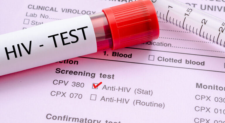
- Rak dojke i laboratorijske pretrage
- Atipična hiperplazija dojke
- Liječenje koštanih metastaza u bolesnica s uznapredovalim rakom dojke
- Nove spoznaje o desenzibilizaciji na antibiotike, kemoterapeutike i biološke lijekove
- Tumorski biljezi (tumorski markeri)
- Toksični učinci antitumorskih lijekova na srce
- Trudnoća nakon liječenja raka dojke
- Hormonska terapija raka dojke
- Mamografija
- Prehrana i rak